CO AND LOW FERRITIN
THE hidden WAY car emissions CAN KILL YOU.
Traffic pollution invades your home—triggering respiratory distress often mistaken for bad indoor air or asthma, while hidden toxins silently block your body’s smallest blood vessels.
Children are among those particularly vulnerable to traffic-related air pollution.

ATP
ATP is the primary energy molecule used by cells. Nearly all cellular functions — including muscle activity, nerve signaling, and repair processes — depend on ATP availability.
Carbon monoxide (CO)
Carbon monoxide is a gas that binds strongly to hemoglobin and interferes with oxygen transport. Even low-level exposure can reduce oxygen delivery to tissues and increase physiological stress.
Cellular energy production
Cellular energy production refers to the processes cells use to generate ATP. Under normal conditions, this mainly occurs through oxygen-dependent mitochondrial respiration.
Chronic hypoxia
Chronic hypoxia means tissues experience long-term reduced oxygen availability. This can force the body into continuous compensatory stress responses and altered metabolism.
Circulation
Circulation is the movement of blood through the body. It delivers oxygen and nutrients to tissues while removing carbon dioxide and waste products.
Compensatory mechanism
A compensatory mechanism is the body’s attempt to maintain stability during dysfunction or stress. These responses may help temporarily, but chronic compensation can eventually contribute to symptoms.
Dysautonomia
Dysautonomia refers to dysfunction of the autonomic nervous system. It can affect heart rate, blood pressure, circulation, digestion, temperature regulation, and stress responses.
Endothelial dysfunction
Endothelial dysfunction means the inner lining of blood vessels no longer regulates circulation normally. This can impair blood flow control, oxygen delivery, vascular relaxation, and microcirculation.
Endothelium
The endothelium is the thin inner lining of blood vessels. It plays a central role in regulating blood flow, vascular tone, inflammation, and oxygen delivery.
Energy deficit
An energy deficit occurs when cells cannot produce enough ATP to meet demand. This can contribute to fatigue, reduced physical capacity, impaired recovery, and physiological stress.
Glycolysis
Glycolysis is a form of energy production that does not require oxygen. It is less efficient than mitochondrial respiration and produces lactate as a byproduct.
Hypoxia
Hypoxia means insufficient oxygen availability at the tissue level. Even when blood oxygen appears normal, impaired circulation can still create localized hypoxia.
Insulin resistance
Insulin resistance occurs when cells respond poorly to insulin signaling. This disrupts glucose regulation and is commonly associated with metabolic dysfunction.
Lactic acid
Lactic acid is produced when cells rely more heavily on glycolysis for energy production. Increased lactic acid production can occur during hypoxia or impaired oxygen utilization.
Lactate
Lactate is a metabolic byproduct generated during glycolysis. Elevated lactate levels may indicate increased reliance on anaerobic energy production.
Metabolic dysfunction
Metabolic dysfunction refers to impaired regulation of energy production and cellular metabolism. It can affect glucose handling, mitochondrial function, circulation, and stress physiology.
Metabolism
Metabolism includes all chemical processes required to maintain cellular function and energy production. It involves how the body uses oxygen, nutrients, and energy.
Microcirculation
Microcirculation refers to blood flow through the body’s smallest vessels, including capillaries. This system is critical for oxygen delivery directly to tissues.
Microvascular dysfunction
Microvascular dysfunction means small blood vessels no longer regulate blood flow properly. This can create uneven oxygen delivery and localized tissue stress.
Mitochondria
Mitochondria are structures inside cells responsible for most ATP production. They generate energy primarily through oxygen-dependent metabolism.
Nitric oxide (NO)
Nitric oxide is a signaling molecule that helps blood vessels relax and regulate circulation. Reduced nitric oxide activity can contribute to vascular dysfunction and impaired blood flow.
Oxidative phosphorylation
Oxidative phosphorylation is the mitochondria’s primary oxygen-dependent energy production pathway. It produces large amounts of ATP under normal physiological conditions.
Oxidative stress
Oxidative stress occurs when reactive oxygen species exceed the body’s antioxidant defenses. Excess oxidative stress can damage cells, blood vessels, and cellular signaling systems.
Oxygen delivery
Oxygen delivery refers to the transport of oxygen from the lungs to tissues. This depends on blood flow, hemoglobin function, vascular regulation, and microcirculation.
Parasympathetic nervous system
The parasympathetic nervous system supports recovery, repair, digestion, and energy conservation. It is often described as the body’s “rest and recovery” system.
Perfusion
Perfusion refers to blood flow reaching tissues and organs. Reduced perfusion can impair oxygen and nutrient delivery even when circulation is still present.
Physiological stress
Physiological stress refers to biological strain placed on the body. This can result from impaired oxygen delivery, metabolic imbalance, inflammation, or chronic compensation.
Redox balance
Redox balance refers to the balance between oxidation and antioxidant defense systems. Disruption of redox balance can contribute to oxidative stress and cellular dysfunction.
Sympathetic nervous system
The sympathetic nervous system activates the body’s stress response. It increases heart rate, blood pressure, alertness, and circulatory compensation during physiological stress.
Tissue hypoxia
Tissue hypoxia means specific tissues are not receiving sufficient oxygen. This may occur even when standard oxygen measurements appear normal.
Vasoconstriction
Vasoconstriction means blood vessels narrow. This can reduce blood flow to tissues and increase circulatory resistance.
Vasodilation
Vasodilation means blood vessels relax and widen. This improves blood flow, oxygen delivery, and tissue perfusion.
The information provided here is for educational purposes only and is not intended as medical advice. Some content discusses hypotheses and proposed biological mechanisms based on an integration of published research. It should not be used to diagnose, treat, cure, or prevent any medical condition. Use this information as a starting point for discussion with a qualified healthcare provider before making decisions about medications, supplements, devices, or changes to your health regimen. See full disclaimer and ai disclosure.
Persistently Low Ferritin as a Marker of Ongoing Carbon Monoxide Exposure
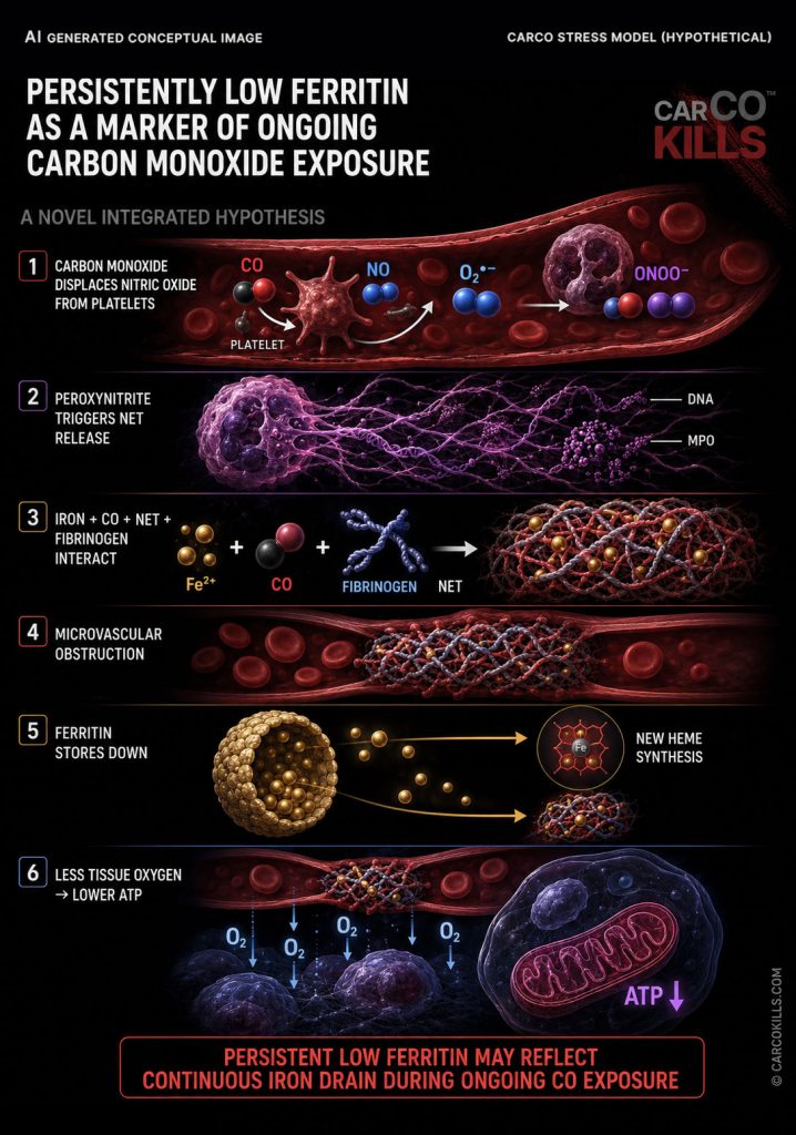
THE CARCO STRESS MODEL: Carbon Monoxide Exposure → Peroxynitrite Formation in Capillaries → Fibrin Amyloid Formation → Iron becomes incorporated into and trapped in fibrin amyloid structures → The iron is not efficiently recycled back into the body’s iron stores → Damaged heme must continuously be replaced → New heme synthesis requires new iron → Iron is repeatedly drawn from ferritin stores → Low Ferritin
Iron Deficiency Without Anemia
Iron deficiency without anemia is a recognized condition where ferritin is low while hemoglobin remains within normal ranges. Symptoms in this state such as fatigue, weakness, reduced physical capacity, exercise intolerance, and impaired recovery are commonly explained as iron deficiency.
Alternative Mechanism in the CARCO Stress Model
The CARCO stress model proposes a different cause for this state of low ferritin. The model indicates that low ferritin is not a true iron deficiency. Instead, persistently low ferritin reflects iron trapping caused by ongoing carbon monoxide exposure.
Low ferritin reflects trapped iron and ongoing carbon monoxide exposure rather than iron deficiency.
Mechanism of Iron Trapping
Repeated carbon monoxide exposure triggers the formation of abnormal fibrin amyloid microclots. Heme-associated iron becomes incorporated into and trapped inside these resistant protein structures during their formation.
Disruption of Normal Iron Recycling
Under normal conditions, iron from degraded heme is recovered and returned to storage. When iron remains trapped in persistent fibrin amyloid structures, it cannot enter the normal recycling pool. This interruption prevents iron from returning to ferritin reserves.
- Carbon monoxide exposure promotes fibrin amyloid formation
- Heme-associated iron becomes trapped in amyloid structures
- Trapped iron is not efficiently recycled
- Damaged heme requires continuous replacement
- New heme synthesis draws iron repeatedly from ferritin stores
- Ferritin stores decline over time
Continuous Heme Replacement Depletes Ferritin
Carbon monoxide and oxidative reactions continuously damage heme-containing structures. Because trapped iron cannot be reused, every newly synthesized replacement heme molecule requires fresh iron drawn directly from existing storage reserves.
Cause of Fatigue and Physical Symptoms
Patients often experience severe fatigue, exercise intolerance, and muscle weakness despite normal hemoglobin levels. In this model, these symptoms result from reduced mitochondrial oxygen availability and cellular energy production failure rather than a lack of iron.
Microvascular Blockage and Energy Loss
Fibrin amyloid structures physically obstruct small blood vessels and reduce blood flow. Mitochondria do not receive enough oxygen to maintain aerobic ATP production, resulting in a persistent cellular energy deficit that limits physical capacity.
Experimental Evidence of Ferritin Reduction During Carbon Monoxide Exposure
Controlled human research has shown that repeated carbon monoxide exposure is associated with a significant reduction in ferritin while hemoglobin mass simultaneously increases.
During a three-week exposure period, ferritin decreased while hemoglobin mass increased. This demonstrates that carbon monoxide exposure can coincide with increased movement of iron out of storage and into heme-containing structures.
The finding provides direct evidence that carbon monoxide exposure can alter iron distribution and reduce ferritin even while hemoglobin production is maintained or increased.
Ferritin Can Recover After Exposure Ends
Carbon monoxide-related changes in iron handling are reversible after exposure ends.
When the processes increasing heme turnover and iron mobilization decline, iron can once again be returned more efficiently to the available iron pool and storage reserves.
Ferritin can therefore recover when the ongoing exposure and the resulting demand for replacement iron stop.
summary
Persistently Low Ferritin as a Marker of Ongoing CO Exposure
Persistently low or repeatedly declining ferritin indicates that iron continues to leave storage faster than it is being restored.
During ongoing carbon monoxide exposure, continued fibrin amyloid formation traps heme-associated iron and reduces its efficient recycling. At the same time, damaged heme continues to require replacement, creating a continuing demand for new iron.
Iron is therefore repeatedly drawn from ferritin stores while less previously used iron returns to replenish them.
Ongoing Carbon Monoxide Exposure → Continued Fibrin Amyloid Formation → Continued Iron Trapping → Reduced Iron Recycling → Continued Heme Replacement → Repeated Withdrawal of Iron From Ferritin Stores → Persistently Low Ferritin
Sources
Schmidt, W. F., Hoffmeister, T., Haupt, S., et al. (2020). Chronic exposure to low-dose carbon monoxide alters hemoglobin mass and V̇O₂max. Medicine & Science in Sports & Exercise, 52(9), 1879–1887. https://pubmed.ncbi.nlm.nih.gov/32118696/
Ghio, A. J., Stonehuerner, J. G., Dailey, L. A., et al. (2008). Carbon monoxide reversibly alters iron homeostasis and respiratory epithelial cell function. American Journal of Respiratory Cell and Molecular Biology, 38(6), 715–723. https://pubmed.ncbi.nlm.nih.gov/18203974/
Rose, J. J., Wang, L., Xu, Q., et al. (2017). Carbon monoxide poisoning: Pathogenesis, management, and future directions of therapy. American Journal of Respiratory and Critical Care Medicine, 195(5), 596–606. https://pubmed.ncbi.nlm.nih.gov/27753502/
Thom, S. R., Fisher, D., & Manevich, Y. (2001). Roles for platelet-activating factor and NO-derived oxidants causing neutrophil adherence after CO poisoning. American Journal of Physiology—Heart and Circulatory Physiology, 281(2), H923–H930. https://pubmed.ncbi.nlm.nih.gov/11454599/
Nielsen, V. G., Cohen, J. B., Malayaman, S. N., Nowak, M., & Vosseller, K. (2011). Fibrinogen is a heme-associated, carbon monoxide sensing molecule: A preliminary report. Blood Coagulation & Fibrinolysis, 22(5), 443–447. https://pubmed.ncbi.nlm.nih.gov/21451399/
Nielsen, V. G., Malayaman, S. N., Khan, E. S., Kirklin, J. K., & George, J. F. (2010). Carbon monoxide releasing molecule-2 increases fibrinogen-dependent coagulation kinetics but does not enhance prothrombin activity. Blood Coagulation & Fibrinolysis, 21(4), 349–353. https://pubmed.ncbi.nlm.nih.gov/20449889/
Pretorius, E., & Kell, D. B. (2014). Diagnostic morphology: Biophysical indicators for iron-driven inflammatory diseases. Integrative Biology, 6(5), 486–510. https://doi.org/10.1039/C4IB00025K
Pretorius, E., Vlok, M., Venter, C., et al. (2021). Persistent clotting protein pathology in Long COVID/Post-Acute Sequelae of COVID-19 (PASC) is accompanied by increased levels of antiplasmin. Cardiovascular Diabetology, 20, 172. https://doi.org/10.1186/s12933-021-01359-7
Everett, J., Brooks, J., Lermyte, F., et al. (2020). Ferritin iron is chemically reduced in the presence of aggregating Aβ(1–42). Scientific Reports, 10, 10332. https://pubmed.ncbi.nlm.nih.gov/32587293/
Nielsen, V. G., Pretorius, E., Bester, J., et al. (2015). Carbon monoxide and iron modulate plasmatic coagulation in Alzheimer’s disease. Current Neurovascular Research, 12(1), 31–39. https://pubmed.ncbi.nlm.nih.gov/25557378/
Kell, D. B., & Pretorius, E. (2014). Serum ferritin is an important inflammatory disease marker, as it is mainly a leakage product from damaged cells. Metallomics, 6(4), 748–773. https://pubmed.ncbi.nlm.nih.gov/24337469/

site updates
The site is continuously updated.
Sources are under review, content will be added to existing topics, and more images will be brought to the site later.
NEW CONTENT COMING:
- News
– Rights to clean air